crystal violet biofilm
Safranin staining provided similar results as crystal violet but with higher reproducibility. Microplates are essential tools for biofilm research since it allows high throughput screening of biofilm forming strains or in the assay of anti-biofilm drugs.

Crystal Violet Quantification Of Biofilm Formation
However biofilm layer formed at the liquid-air interphase known as pellicle is extremely sensitive to its washing and staining steps.

. In this method the negatively charged molecules within a. Reagent reservoirs if using a multichannel pipette. - Stain with 1 of Crystal violet.
30 vv glacial acetic acid solution. Early phase biofilms are also prone to damage by the latter steps. With immature biofilms Figure 1A individual cells can be distinguished and counted.
Adherent biofilms were fixed for 1 h at 60 C stained by 200 µl Hucker modified crystal violet Sigma Chemical Company- USA for 5 min at room temperature and then rinsed with water and allowed to dry. As edge effect causes a significant increase in plate. Distilled sterile water for washing.
01 wv Crystal Violet solution. In the former biofilms are stained with CV and distained with a decolorization solution that can be measured spectrophotometrically. Cover assay plates and incubate at optimal growth temperature for desired amount of time.
The crystal violet assay is widely used for biofilm quantitation despite its toxicity and variability. Biofilm biomass and metabolic activity were quantified with crystal violet and methyl thiazolyl tetrazolium staining methods. Incubator 37C for 15 min then air-dry for 15 min.
Following incubation planktonic bacteria are rinsed away and the remaining adherent bacteria biofilms are stained with crystal violet dye thus allowing visualization of the biofilm. 3 Set up four small trays in a series and add 1 to 2 inches of tap water to the last three. Biofilm samples were destained with.
Images of Pseudomonas aeruginosa biofilms stained with crystal violet at different incubation times are shown in Figure 1. In this assay bacteria are incubated in vinyl U-bottom or other types of 96-well microtiter plates. 1- grow the bacteria 2- guarantee the pure culture 3- next form the biofime add 500 microliters of bacteria into a 24 well microplate.
1 Paytubi et al 2017. Merritt et al 2005. This method allows for the in vitrocultivation and quantification of bacterial biofilms12The CV.
Micropipettes pipettes and polystyrene macro cuvette. Here we instead combine fluorescence labelling with the Cytation 5 multi-mode plate reader to enable simultaneous acquisition of both quantitative and imaging biofilm data. However most isolates in.
Rings of crystal violet around a well are not indicative of biofilm formation and should be rinsed again as excess stain will skew the results of the assay. Leave the plate face up on the bench top overnight to dry. One of the first staining assays used in biofilm analysis was the crystal violet assay.
However 96 well microtitre plate based assays share the issue of edge effect. Allow the acetic acid to. Crystal violet CV assay is the most popular method for biofilm determination adopted by different laboratories so far.
A variety of methods have been developed to quantify biofilm formation including crystal violet CV staining and XTT reduction assay XTT 23 24. This can be time consuming require many images for reproducibility and be subject to user bias as mentioned with colony-counting. Crystal violet assay was performed to assess the biofilm forming abilities based on optical density obtained.
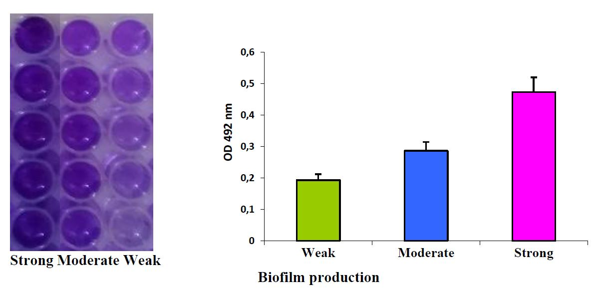
Based on adherence strength the biofilm forming abilities were classified into four different categories. Assessment of the total biomass by crystal violet CV staining assay CV is used in Gram staining to differentiate between Gram-positive and -negative bacteria and is the most common staining method to measure the total biomass of sessile cells in both ALIM and MPM Figs. 1 Phosphate-buffered saline PBS.
4- keep without no agitation for 24 or 48 or 72 days until. Multichannel micropipette 20200 μl volume and sterile tips see Note 4. - Shaking out the liquid wash one time with 200 ul dH2O and pipet out slowly to avoid disrupt the biofilm.
Inoculate biofilm assay plates directly in 100-μl medium per well from the overnight microtiter plate cultures using a sterile 96-prong inoculating manifold. In order to quantify the biofilm production capabilities of an isolate the Crystal Violet CV assay is often preferred due to its simplicity reliability and quick throughput. 22 Materials for Crystal Violet Biofilm Staining and Detection 1.
Aureus biofilm formation the crystal violet CV assay and the XTT tetrazolium salt reduction assay were optimized evaluated and further compared. Quantification of Biofilms by Crystal Violet Staining Assay 1. Aureus biofilm was sensitive to food heat processing conditions with.
Crystal violet staining is commonly used for quantification of biofilm formation although it is highly toxic. To measure biofilm formation biofilms were rinsed three times with distilled water incubated with 01 g v crystal violet for 20 min at room temperature and solubilized in 95 ethanol after removing crystal violet and washing with distilled water three times. Here we test safranin as a non-toxic replacement.
2 5 7 and 8 and Table. To prepare the solution the required quantities were diluted in sterile distilled water. The primary cause of the edge effect phenomenon is evaporation.
Non-adherent weakly adherent moderately adherent and. Add 200 μL of 30 acetic acid to all wells that were stained to solubilize the crystal violet. Two assays for quantification of S.
We therefore recommend safranin staining for biofilm biomass quantification. 05 wv crystal violet solution in deionized water. Correlation of crystal violet biofilm test results of Staphylococcus aureusclinical isolates with Raman spectroscopic read-out Christina Ebert Christina Ebert Leibniz Institute of Photonic Technology Jena Germany Center for Sepsis Control and Care Jena University Hospital Jena Germany Search for more papers by this author Lorena Tuchscherr.
In CV assay most isolates formed weak biofilm 743 while the rest formed moderate biofilm 233 or strong biofilm 23.

Quantification Of Biofilm Formation By Crystal Violet Staining Download Scientific Diagram

Biofilm Formation Assays In Regulated Polyp Deficient Pseudomonas Sp Download Scientific Diagram

Biofilm Formation Assay Kit Testpiece Dojindo Eu
Study On The Promotion Of Bacterial Biofilm Formation By A Salmonella Conjugative Plasmid And The Underlying Mechanism Plos One
Ijms Free Full Text Expression Of The Biofilm Associated Genes In Methicillin Resistant Staphylococcus Aureus In Biofilm And Planktonic Conditions Html

A The Crystal Violet Staining Results Of Biofilms After Culturing Download Scientific Diagram

Crystal Violet Assay To Assess The Antibiofilm Activity Of Samples Download Scientific Diagram

Schematic Crystal Violet Assay On Biofilms In A Microtiter Plate Download Scientific Diagram

Crystal Violet Assay Crystal Violet Was Used To Stain Biofilm Download Scientific Diagram

Eal And Rpff Contribute To Biofilm Formation A Crystal Violet Download Scientific Diagram

A Crystal Violet Cv Staining Of Biofilms Formed By Pao1 Under Download Scientific Diagram

Role Of Camp In Biofilm Formation A Crystal Violet Staining Of Download Scientific Diagram
Quantification Of Biofilm Formation By Crystal Violet Staining Download Scientific Diagram

Figure 2 Investigation Of Biofilm Forming Ability In Staphylococci Causing Bovine Mastitis Using Phenotypic And Genotypic Assays

Crystal Violet Staining Was Performed To Assess Air Liquid Biofilm Download Scientific Diagram

Screening Of Biofilm Formation With Crystal Violet Staining By The 96 Download Scientific Diagram

Production Of Biofilm Was Detected By Crystal Violet Staining A And Download Scientific Diagram

Biofilm Assay On Various Polystyrene Surfaces Crystal Violet Biofilm Download Scientific Diagram

Pdf An Improved Crystal Violet Assay For Biofilm Quantification In 96 Well Micro Titre Plate


Comments
Post a Comment